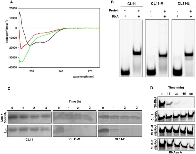
Fig. 3.
Characterization of CL11, CL11-M, and CL11-E variants and their binding to 58rRNA. (A) CD spectra of CL11 (black), CL11-M (red), and CL11-E (green) in buffer R. (B) EMSA assay where equimolar concentration of f58rRNA target was incubated with the different protein variants. Free f58rRNA was used as a negative control. (C) Proteolytic digestion of CL11, CL11-M, and CL11-E by Lon protease. The proteins were incubated with Lon in the presence or absence of 58rRNA in the reaction mixture. (D) f58rRNA digestion by RNase A in presence or absence of CL11, CL11-M, and CL11-E protein in the Lon buffer at 37°C.

