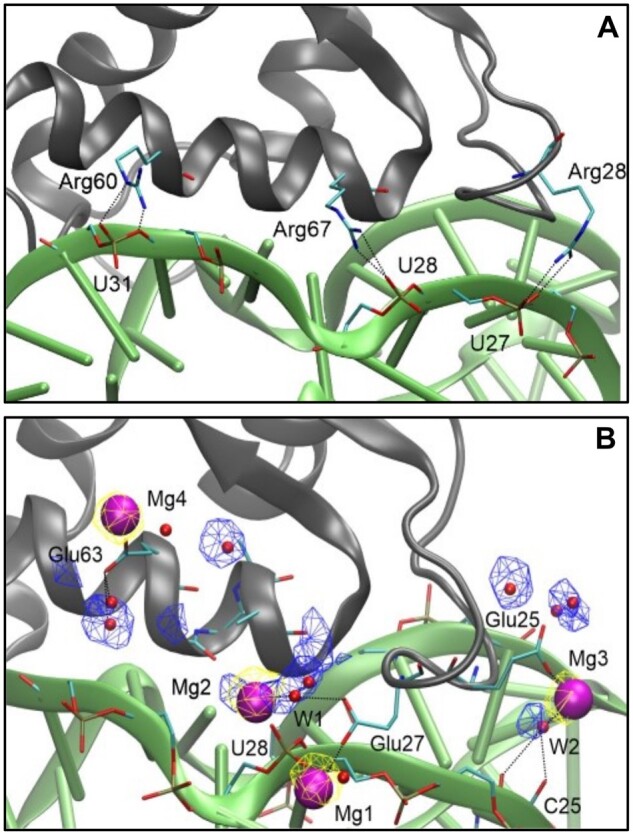
Fig. 5.

Molecular details of RNA–protein interaction. Representative snapshots from the last 500 ns of MD simulations showing the CL11/CL11-E proteins (gray cartoon) with important residues in sticks (cyan: C, blue: N, red: O, golden: P, hydrogens not shown), 58rRNA (green cartoon) with important residues in sticks, Mg2+ ions (purple spheres), and water molecules (red spheres). Yellow and blue meshes show the conserved sites for Mg2+ and water, respectively, at 0.2 and 0.25 occupancy isovalue. Black dotted lines indicate H-bonding or metal coordination. (A) CL11–58eRNA, (B) CL11-E–58rRNA.
