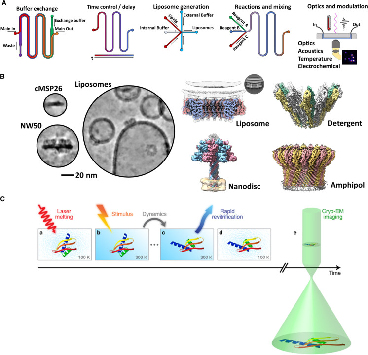
Figure 2. Static and dynamic processes elucidated by cryoEM.
(A) Microfluidic devices enable complex biochemistry and processes to be performed and studied on-chip. These chips also enable rapid, time-resolved cryoEM. Shown from left to right: microfluidic buffer exchange, time delay for temporal control, liposome generation, mixing of reactions and last, the coupling of detectors/optics to a microfluidic chip for data collection. (B) Membrane mimetic technologies enable structural studies of PFPs in a near-native lipid membrane context. Shown are various examples of nanodiscs [15,161–163], liposomes [39,163], detergent [4] and amphipols [88,164] which have all been used to study the pore and prepore forms of PFPs. Select cryoEM examples; MPEG1 [39], XaxAB [4], Anthrax toxin [15] and polyC9 [88] (C) Microfluidic and in situ time-resolved cryoEM techniques provide structural insight over small increments of time. Panel C reproduced from Voss et al. [90]. Stimulation by laser (red) melts the vitreous ice, enabling rapid (photo-dependent; ‘stimulus') conformational changes to occur before re-vitrification. Followed by standard cryoEM imaging (green).

