Figure 6.
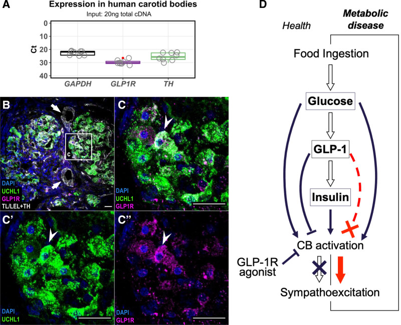
GLP1 (glucagon-like peptide 1) receptor expression in human carotid bodies. A, GLP1R mRNA expression in human carotid bodies (CBs) assessed by RT-qPCR. Expression presented as Ct values in comparison to housekeeping (GAPDH) and reference (TH) genes. All reactions were performed on the same plate. TH – Tyrosine Hydroxylase; Boxplot hinges represent interquartile range (IQR=Q3–Q1). Red dot indicates an outlier (Q3+1.5*IQR). n=5. B and C, Localization of GLP1R in human CBs. GLP1R immunoreactivity (magenta) was detected in chemosensory glomus cells marked by UCHL1 (green). Arrows indicate blood vessels devoid of GLP1R immunoreactivity. Arrowhead indicates GLP1R-positive chemosensory glomus cell. n=1. Representative image selected best demonstrating GLP1R localization in a well-defined chemosensory glomus cell cluster. Scale bar—20 µm. D, Schematic model of GLP1 action on CBs in regulating sympathetic activity. Upon food ingestions, rise in blood glucose activates the CBs leading to sympathoexcitation and stimulates release of GLP1 from intestinal L-cells. GLP1 mediates insulin secretion which additively stimulates the CB chemoreceptors. GLP1 inhibits the chemosensory CB cells counteracting the sympathoexcitation to elevated levels of glucose and/or insulin in normal physiological context. Disruption of the GLP1 inhibitory component leads to aberrant sympathoexcitation as shown in the SH model. Treatment with GLP1R agonists act to reduce sympathetic activity by suppressing the peripheral chemoreflex drive from the CBs. TL/LEL indicates tomato lectin/lycopersicon esculentum lectin; and UCHL1, ubiquitin carboxy-terminal hydrolase L1.

