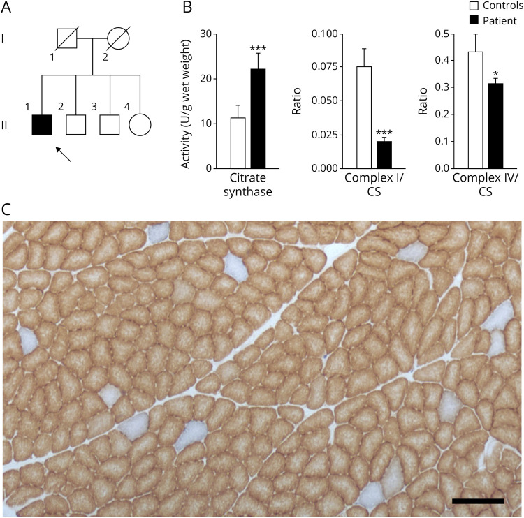
Figure 1. Mosaic Mitochondrial Impairment in Skeletal Muscle of a Patient Having Neuropathy, Ataxia, and Retinitis Pigmentosa Syndrome.
(A) Pedigree of the patient's family. The patient is indicated by an arrow. (B) Spectrophotometric measurements of mitochondrial enzyme activities in skeletal muscle homogenate of the patient and controls. NADH:ubiquinone oxidoreductase (complex I) and cytochrome c oxidase (complex IV) activities were normalized to citrate synthase activities. Mean values of 3 independent measurements are shown for the patient, whereas control values indicate averages of 3 healthy individuals. Error bars represent standard deviations. For significance, the t test was performed; *p < 0.05; ***p < 0.001. (C) COX/SDH double staining of the patient's skeletal muscle. Brown color indicates functional COX, whereas blue color results from SDH activity in the absence of COX activity. Scale bar, 200 µm. COX = cytochrome c oxidase; SDH = succinate dehydrogenase.

