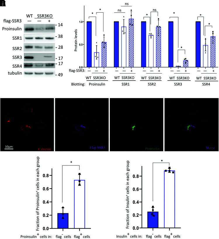
Figure 5.
TRAPγ/SSR3 re-expression rescues proinsulin protein levels in TRAPγ/SSR3-KO β-cells. TRAPγ/SSR3-KO INS832/13 cells were transfected with empty vector (−) or plasmids encoding Flag-tagged TRAPγ/SSR3 cDNA (+). Wild-type (WT) INS832/13 cells were also transfected with empty vector as control. A: At 48 h posttransfection, cells were lysed and analyzed by reducing SDS-PAGE and immunoblotting for anti-rodent proinsulin and the additionally indicated antibodies. B: Quantitation (mean ± SD) of proinsulin and each TRAP/SSR subunit from four independent experiments like that shown in A; expression levels in WT control cells were set to 1.0. C: TRAPγ/SSR3-KO INS832/13 cells transfected as in A were examined by immunofluorescence with anticalnexin (red), anti-Flag (Flag-SSR3, purple), and anti-rodent proinsulin (green); a merged anti-Flag/antiproinsulin image is shown. D: Quantitation (mean ± SD) of rodent proinsulin–positive TRAPγ/SSR3-KO cells that either re-express or do not re-express TRAPγ/SSR3 protein from three independent experiments like that shown in C. E: Quantitation (mean ± SD) of insulin-positive TRAPγ/SSR3-KO cells that either re-express or do not re-express TRAPγ/SSR3 protein from four independent experiments. *P < 0.05.

