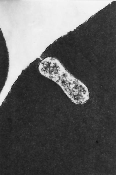
FIG. 4.
Electron photomicrograph of intraerythrocytic B. henselae, illustrating the existence of a pore between the bacterium and the extracellular fluid space. Sample was stained with methanol, uranyl acetate, and lead citrate. Magnification, ×38,000. (Reproduced from reference 55 with permission of the publisher.)

