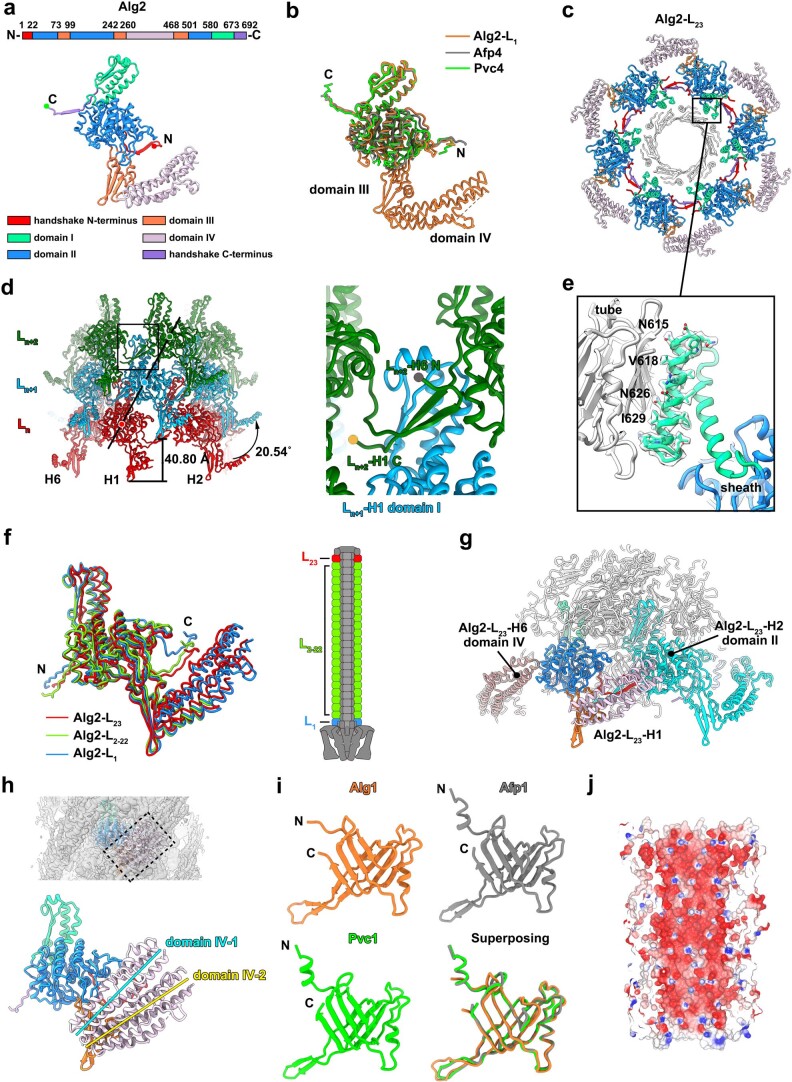
Extended Data Fig. 5. Structural analyses of sheath-tube module in extended state.
a: Schematic and ribbon diagrams showing that the sheath protein (Alg2) has four domains. The N- and C-termini are marked with red and green circles. b: Structural superpositions of sheath protein (Alg2: orange) with homologous proteins (Pvc4: green; Afp4: grey). The additional domains (domain III and IV), N-, and C-termini of Alg2 are labeled. c: Ribbon diagram of a perpendicular slice of the distal sheath layer (Alg2-L23), showing the domain organization of Alg2 in one sheath layer and the interactions between the sheath and inner tube (white). The color code matches panel a. Box indicates the interface between sheath and inner tube, which is shown in panel e. d: Ribbon diagrams showing an extended sheath fragment containing three sheath layers (Ln, Ln+1 and Ln+2) in different colors. The direction of one helical strand (H1) is highlighted by a line. Box indicates the conserved handshake interaction between sheath subunits, which is shown in the right panel. e: Shadowed surface and ribbon diagram showing the interactions between the attachment helix of sheath and inner tube. The residues mediating contacts are labeled and shown with side chains. f: Structural superpositions of sheath subunits from different layers (distal layer: red; central layers: green; proximal layer: blue) showing the structural diversity in domain IV, N-, and C-termini of Alg2 across different sheath layers. The schematic (right) indicates the position of the different sheath layers on the AlgoCIS particle. g: Side view of ribbon diagram showing the overall structure of the cap module and the distal sheath layer. The inner tube proteins, cap, and cap adaptor are colored white. The color code for different domains of one Alg2 subunit (Alg2-L23-H1) matches panel a, whereas the neighboring two subunits are shown in different colors (Alg2-L23-H2: cyan, Alg2-L23-H6: brown). h: Shadowed surface and ribbon diagrams showing that there are two conformations of domain IV (domain IV-1, 2) in the central sheath layers. The color code for different domains matches panel a, while two conformations of domain IV are represented by lines with different colors (domain IV-1: blue; domain IV-2: yellow). i: Ribbon diagrams showing the structures of inner tube protein (Alg1: orange) and the homologs (Afp1: grey; Pvc1: green). The structural superpositions of Alg1 with homologs are shown in the bottom right panel. j: Surface electrostatic potential of the inner tube showing that the inner surface of tube is dominated by negative charges. Negative and positive electrostatic potentials are colored red and blue, respectively.

