Fig. 5.
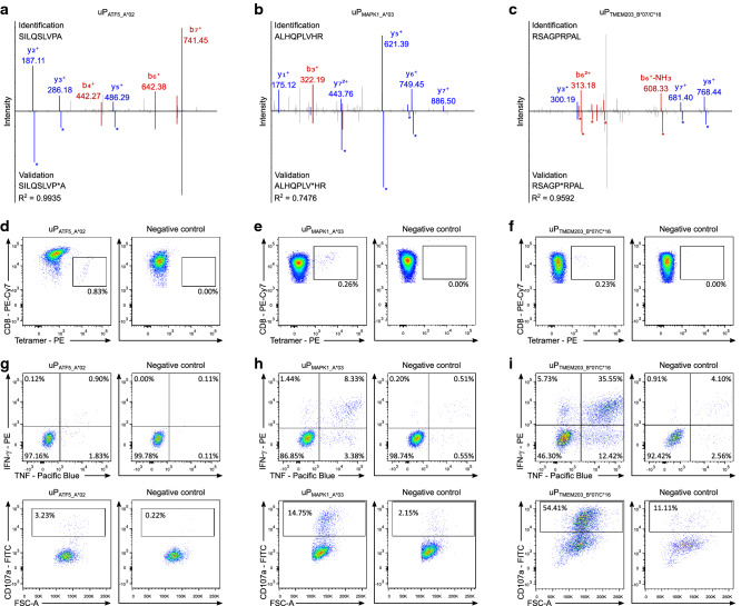
Spectral validation and immunogenicity analyses of uORF-derived tumor antigens. a–c Validation of the experimentally eluted peptides a uPATF5_A*02, b uPMAPK1_A*03, and c uPTMEM203_B*07/C*16. Comparison of fragment spectra (m/z on x-axis) of HLA uLigands eluted from primary samples (identification) to their corresponding isotope-labeled synthetic peptides (validation, mirrored on the x-axis) with the calculated spectral correlation coefficient (R2). Identified b- and y-ions are marked in red and blue, respectively. Ions containing isotope-labeled amino acids are marked with asterisks. d–f Naïve CD8+ T cells were primed in vitro using HLA uLigand-loaded aAPCs with the HLA-A*02-, -A*03-, and -B*07-restricted peptides d uPATF5_A*02, e uPMAPK1_A*03, and f uPTMEM203_B*07/C*16, respectively. Graphs show single, viable cells stained for CD8 and PE-conjugated multimers of indicated specificity. The left panels show HLA uLigand tetramer staining, the right panels (negative control) depict tetramer staining of T cells from the same donor primed with an HLA-matched control peptide. g–i Functional characterization of HLA uLigand-specific CD8+ T cells by intracellular cytokine staining. Representative examples of IFN-γ and TNF production (upper panels) as well as CD107a expression (lower panels) after stimulation of g uPATF5_A*02-, h uPMAPK1_A*03-, and i uPTMEM203_B*07/C*16-specific CD8+ T cells with the HLA-A*02-, -A*03-, and -B*07-restricted peptides uPATF5_A*02, uPMAPK1_A*03, and uPTMEM203_B*07/C*16, respectively (left panels) compared to a negative HLA-matched control peptide (right panels)

