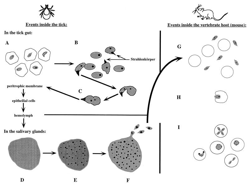
FIG. 1.
Life cycle of Babesia spp. in the tick and vertebrate hosts. Events in the tick begin with the parasites still visible in consumed erythrocytes. Some are beginning to develop Strahlenkörper forms (A). The released gametes begin to fuse (note that only one of the proposed mechanisms is pictured; one gamete has a Strahlenkörper form, whereas the other does not) (B). The formed zygote then goes on to infect and move through other tissues within the tick (C) to the salivary glands. Once a parasite has infected the salivary acini, a multinucleate but undifferentiated sporoblast is formed (D). After the tick begins to feed, the specialized organelles of the future sporozoites form (E). Finally, mature sporozoites bud off of the sporoblast (F). As the tick feeds on a vertebrate host, these sporozoites are inoculated into the host (G). Not shown is the preerythrocytic phase seen in Theileria spp. and T. equi (B. equi). Sporozoites (or merozoites) contact a host erythrocytic and begin the process of infection by invagination (H). The parasites become trophozoites and can divide by binary fission within the host erythrocyte, creating the various ring forms and crosses seen on stained blood smears (I). Illustrations are not to scale.

