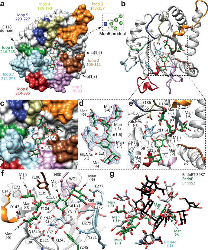
Fig. 8. Structural basis of EndoE GH18 domain substrate specificity.
a Surface representation of the EndoE-GH18L-Man5 crystal structure, with annotated domains and loops. b Cartoon representation of the EndoE-GH18L-Man5 crystal structure. c Surface representation of the GH18 domain of EndoE showing the location of the Man5 product into the active site. d Electron density map of the Man5 product shown at 1.0 σ r.m.s.d. e Cartoon representation of the GH18 domain of EndoE showing the location of the Man5 product into the active site. f Cartoon representation of the GH18 domain of EndoE showing the main residues and secondary structure elements interacting with the Man5 product in the active site. g Superposition of the HM-type N-glycan found in the X-ray crystal structures of EndoE-GH18L (green), EndoBT-3987 (black), and EndoS2 (gray).

