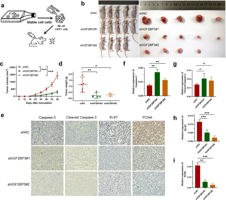
Fig. 3. Knockdown of IGF2BP3 decreases AML cell viability in vivo.
a Schematic diagram showing the schedule of nude mouse xenograft assays. b Subcutaneous tumors were observed at 36 days in two different groups (the black arrows indicate xenograft tumors). c The xenograft growth curves for the shIGF2BP3#1, shIGF2BP3#2, and shNC groups were plotted by measuring the tumor size (width2 × length × π/6) with a Vernier caliper every four days. d Nude mice were sacrificed, and xenografts were harvested and weighed. e–i Representative images of immunohistochemical staining for Caspase-3, cleaved Caspase-3, Ki-67, and PCNA in tumors excised from xenograft model mice. *P < 0.05; **P < 0.01; ***P < 0.001; ns, not significant.

