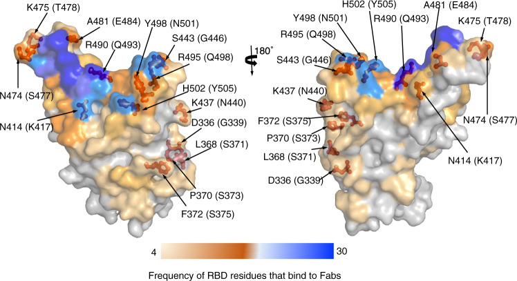
Fig. 4. Mutations in the omicron spike RBD that potentially allow the omicron variant to escape from known neutralizing antibodies.
Forty-nine PDBs of neutralizing antibody/RBD complexes were analyzed using PDBePISA (https://www.ebi.ac.uk/pdbe/pisa/). RBD residues are colored based on their frequency in interacting with known neutralizing antibodies: from light orange (low frequency) to blue (high frequency). RBD residues that have undergone mutations in the omicron RBD are shown as red sticks. Structural data for neutralizing antibody/RBD complexes were obtained from the PDB: 6wpt, 6xc2, 6xc4, 6xcm, 6xdg, 6xkp, 6xkq, 6yor, 7a5r, 7akd, 7b3o, 7bwj, 7byr, 7c01, 7cac, 7cdi, 7cdj, 7ch4, 7ch5, 7chb, 7chh, 7cho, 7chp, 7chs, 7cjf, 7eam, 7ean, 7jx3, 7k8m, 7k8v, 7k8w, 7k8x, 7k43, 7lrs, 7m6d, 7m6f, 7m6g, 7m6h, 7m7w, 7m42, 7mkl, 7mlz, 7n4i, 7n4j, 7n4l, 7n4m, 7r6w, 7r7n, 7sn2. Fab: antigen-binding fragment.

