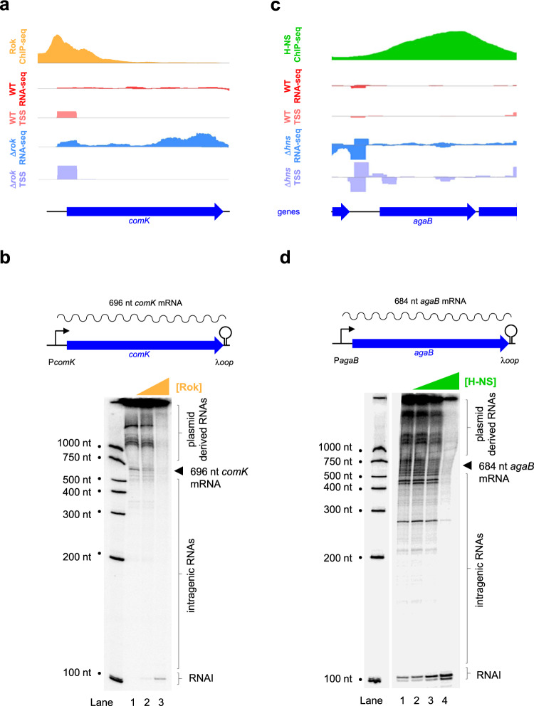
Fig. 2. Housekeeping RNA polymerases of E. coli and B. subtilis differ in their promiscuity.
a Rok represses transcription of the B. subtilis comK mRNA in vivo. Data for Rok occupancy (orange)35, total RNA abundance (red and blue) and transcription start site (TSS) usage (pink and mauve) are shown. Sequence reads mapping to the top and bottom DNA strands are shown above and below the central horizontal line in each plot. The y-axis scales are identical for data obtained using wild-type and Δrok cells for each type of experiment. Genes are shown by blue arrows. b Rok represses transcription of the B. subtilis comK mRNA in vivo. The schematic illustrates the comK gene and regulatory region, cloned in plasmid pSR, and used as a template for in vitro transcription. The comK TSS is shown as a bent black arrow, the comK gene is shown as a block blue arrow, and the sequence encoding the λoop transcriptional terminator is indicated by a stem loop schematic. B. subtilis σA RNA polymerase (0.5 μM) and Rok (0, 0.5, or 1.0 μM) were added as indicated. Note that the 696 nt comK mRNA is easily discernible and there is no evidence for transcription initiation within comK. Species of RNA over ~1000 nt in length are derived from sites elsewhere on the plasmid template. The RNAI transcript is encoded by the plasmid replication origin. The experiment was done twice with similar results. c H-NS represses transcription initiation within the E. coli agaB coding sequence in vivo. Data for H-NS occupancy are in green32 and otherwise as indicated in (a) except that wild-type and Δhns E. coli cells are compared. d H-NS represses transcription initiation within the E. coli agaB coding sequence in vitro. The schematic illustrates a section of DNA cloned in plasmid pSR and used as a template for in vitro transcription. The expected size of the agaB mRNA is 684 nucleotides (nt). The gel image shows transcripts generated by E. coli σ70 RNA polymerase (0.5 μM) using this DNA template. The expected position of agaB mRNA is indicated by an arrow head but is obscured by many similarly sized and smaller transcripts derived from agaB coding sequence. H-NS was added at concentrations of 0, 0.5, 1.0 or 2.0 μM. The experiment was done twice with similar results.

