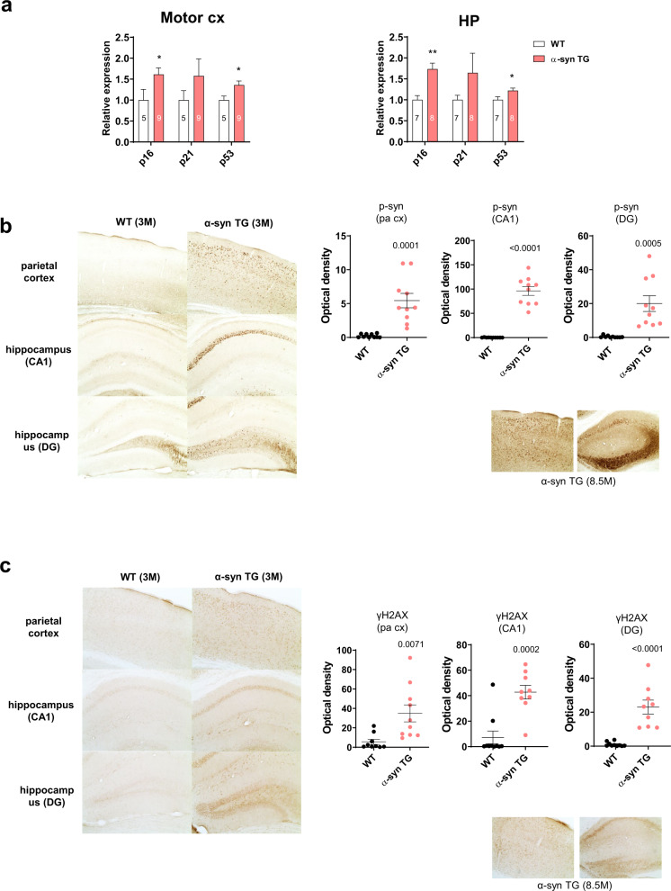
Fig. 7. DSB changes in α-synuclein tg mice.
a p16, p21, and p53 mRNA levels increased in the motor cortex (cx) and hippocampus (HP) in α-syn tg mice compared with those of wild-type mice. b Immunohistochemistry of WT and α-syn tg (3 months) mice with the anti-phospho-α-synuclein antibody. Note that phosphorylated α-synuclein increased in the parietal cortex, hippocampal CA1 region, and DG of α-synuclein tg mice. Phosphorylated α-synuclein increased more in the older tg (8.5 months) mice than in the 3-month tg mice. c Immunohistochemistry of WT and α-syn TG (3 months) mice with the anti-γH2AX antibody. Note that γH2AX increased in the parietal cortex, hippocampal CA1 region, and DG of α-synuclein tg mice. γH2AX increased more in the older tg (8.5 months) mice than in the 3-month tg mice.

