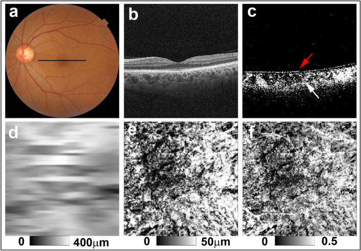
Figure 2.
PS-OCT imaging of the left eye of a 56-year-old woman from the non-sunset group (Case 4 in Supplementary Table S1). (a) Color fundus images showed no evidence of sunset glow fundus. The black line in the color fundus image designates the scan line for PS-OCT B-scan images. (b) Standard OCT B-scan image. (c) Binary DOPU B-scan image (DOPU < 0.8) showing preservation of RPE melanin (red arrow) and choroidal melanin (white arrow). (d) Choroidal thickness map, (e) ChMeT map, and (f) ChMeTratio map.

