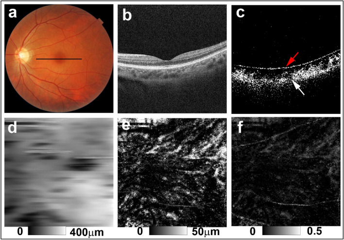
Figure 3.
PS-OCT imaging of the left eye of a 43-year-old woman from the potential sunset group (Case 6 in Supplementary Table S1). (a) Color fundus images showed faint diffuse depigmentation. The black line in the color fundus image designates the scan line for PS-OCT B-scan images. (b) Standard OCT B-scan image. (c) Binary DOPU B-scan image (DOPU < 0.8) showing preservation of RPE melanin (red arrow) and mild reduction of choroidal melanin (white arrow). (d) Choroidal thickness map. (e) ChMeT map and (f) ChMeTratio map showing decreased choroidal melanin compared with healthy control eyes (Fig. 1) and non-sunset eyes (Fig. 2).

