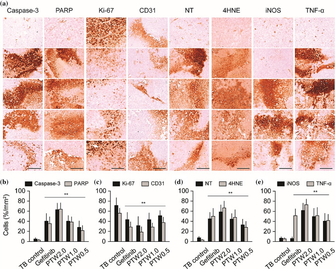
Fig. 32.
Immunohistochemistry in tumor mass. Tumor tissue was stained for caspase-3, PARP for apoptosis, Ki-67 for cell proliferation, CD31 for angiogenesis, nitrotyrosine (NT) and 4-hydroxynonenal (4HNE) for oxidative stress, and iNOS, TNF-α for immune response (A). The percentages of immune-reactive cells are shown in (B–E) [178]

