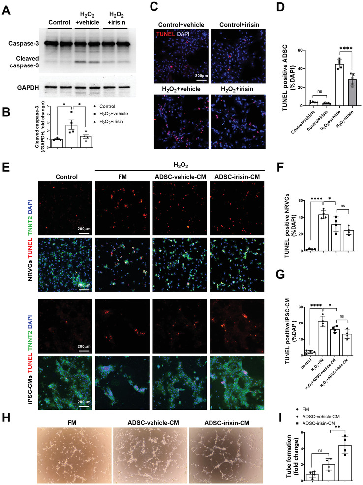
Figure 5.

Irisin pretreatment protects ADSCs against apoptosis and enhances their ability to increase angiogenesis. A,B) Western blots and quantification of cleaved caspase‐3 protein expression in ADSCs. ADSCs were pretreated with or without irisin (100 ng mL−1, 48 h), washed to remove irisin, and then subjected to H2O2 (200 µm, 6 h). C) Representative images of TUNEL‐positive ADSCs (red) with or without H2O2 (200 µm, 6 h). Cell nuclei were stained with DAPI (blue). D) Quantification of TUNEL positive ADSCs (n = 5). E–G) Representative images of TUNEL‐positive (red) NRVCs and iPSC‐CMs. NRVCs and iPSC‐CMs were treated with FM, ADSC‐vehicle‐CM, or ADSC‐irisin‐CM 15 min before H2O2 (200 µm, 6 h) administration. NRVCs and iPSC‐CMs are TNNT2 positive (green). n = 4. H,I) The paracrine angiogenesis of ADSC‐vehicle‐CM and ADSC‐irisin‐CM was evaluated by rCAEC tube formation assays. n = 4. The data were analyzed by 1‐way ANOVA, followed by a Bonferroni post hoc test. *p < 0.05, **p < 0.01, ****p < 0.0001; ns, not significant.
