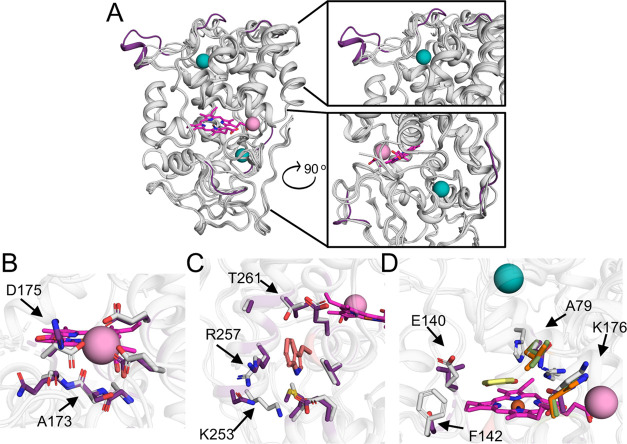
Figure 4.
Structural basis for the different activity profiles in VPs. (A) AlphaFold223 models of 5H, 8H, and 11H are superimposed onto the VPL crystallographic structure (PDB entry 3FJW). All VP backbones are presented in gray cartoons, VPL calcium and manganese ions are in teal and pink spheres, respectively, and the heme group in pink sticks in all panels. Backbone segments that are unique for 8H are colored in purple. (B) Manganese-oxidation site. VPL and 8H residues that chelate manganese and vicinal side chains are in gray and purple sticks, respectively. Significant variations are marked in arrows and their position identities and numbers are relative to the PDB entry 3FJW in all panels. (C) Reactive surface tryptophanyl site. Tryptophan is presented in salmon sticks. VPL and 8H residues in the tryptophan vicinity are presented in gray and purple sticks, respectively. (D) Access channel to the heme-oxidation site. Guaiacol (GUA), which is chemically similar to DMP, from a VPL crystallographic structure (PDB entry 4G05(32)) is in yellow sticks. VPL residues in the GUA vicinity are gray. Mutations relative to VPL are indicated in green, purple, and orange sticks for 5H, 8H, and 11H, respectively, and are marked by arrows.

