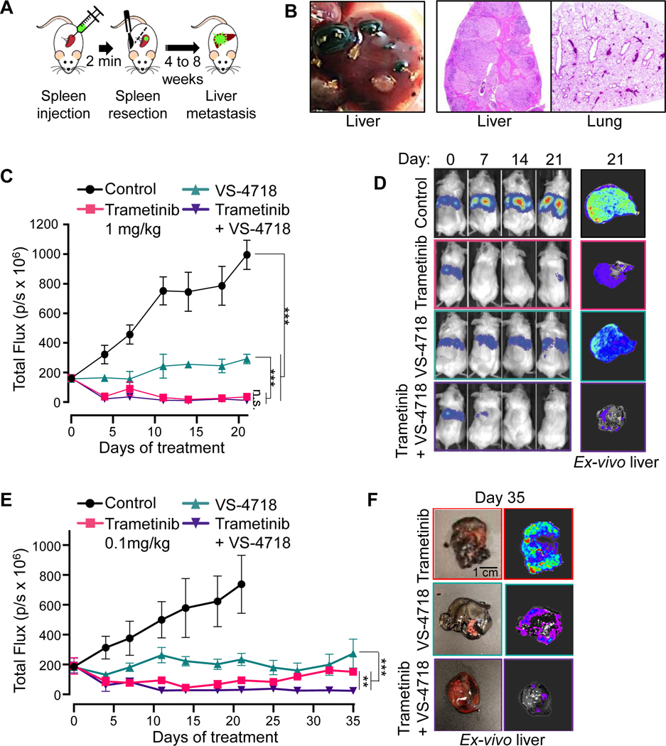
Figure 5. MEKi/FAKi combination reduces UM cells growth in an in vivo liver metastasis model.
(A) Schematic of the hematogenous dissemination model for UM liver metastasis using 92.1 GFP-Luc cells. (B) Left, Macroscopic view of liver metastasis 8 weeks post-splenic injection. Right, H&E staining of liver and lung. (C) Hepatic tumor burden tracked by IVIS imaging after injection of 92.1 UM cells in SCID/NOD mice treated with vehicle (Control), trametinib 1 mg/kg, VS-4718 50 mg/kg or both. Data are mean±SEM (6 mice/group). ***p<0.001; n.s. not significant. (D) Representative mice treated with vehicle (Control), trametinib 1 mg/kg, VS-4718 50 mg/kg or both, at the indicated days of treatment, and representative ex-vivo imaging of the liver obtained at day 21. (E) Hepatic tumor burden tracked by IVIS imaging after injection of 92.1 UM cells in SCID/NOD mice treated with vehicle (Control), trametinib 0.1 mg/kg, VS-4718 50 mg/kg or both. Data are mean±SEM (5 mice/group). ***p<0.001; **p<0.01. (F) Representative ex-vivo imaging of the liver from mice treated for 35 days with trametinib 0.1 mg/kg, VS-4718 50 mg/kg or both.

