
Figure 4.
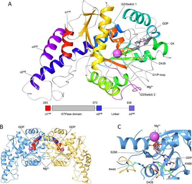
Structural organization of OPA1-MGD monomer and dimer. A) The three-dimensional structure of GDP-bound OPA1-MGD monomer is shown as cartoon, secondary structure elements are colored in a red-to-blue scheme according to the sequence, GDP is shown as sticks with C atoms in grey, O atoms in red, N atoms in blue and P atoms in orange, Mg2+-ion is represented as a pink sphere, residue D438 is shown as sticks with C atoms in yellow, N atoms in blue and O atoms in red. Functional element G1/P-loop is colored in yellow, G2/Switch 1 in cyan, G3/Switch 2 in magenta, G4 in dark grey. Inset shows the structural organization of OPA1-MGD with the three helices of the Helix Bundle colored as above. B) The three-dimensional structure of GDP-bound OPA1-MGD dimer is represented as cartoon with protomers in blue and yellow, Mg2+-ion is represented as a pink sphere, GDP is shown as spheres with C atoms in grey, N atoms in blue, O atoms in red, P atoms in orange. C) Intra-intermolecular interactions involving D438 residue. The three-dimensional structure of GDP-bound OPA1-MGD dimer is represented as cartoon with protomers in blue and yellow green, Mg2+-ion is represented as a pink sphere, GDP, S298, D438, R445 and K468 are shown as sticks with C atoms in grey, dark teal, yellow, cyan, and light green respectively, N atoms in blue, O atoms in red, P atoms in orange. H-bonds are represented by yellow dashed lines, salt bridges by magenta dashed lines.