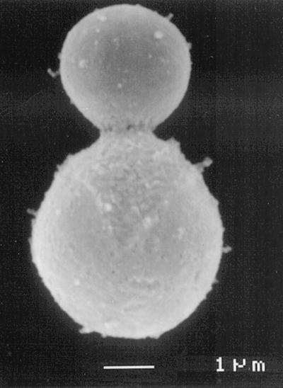
FIG. 2.
Electron micrograph of a “melanin ghost” of C. neoformans. Organs from a mouse infected with Mel+ C. neoformans were extracted with solvents, digested in hot HCl, and centrifuged. These structures were not seen when a Mel− infection was so treated. Courtesy of Rosas et al. (For a related study, see reference 71).

