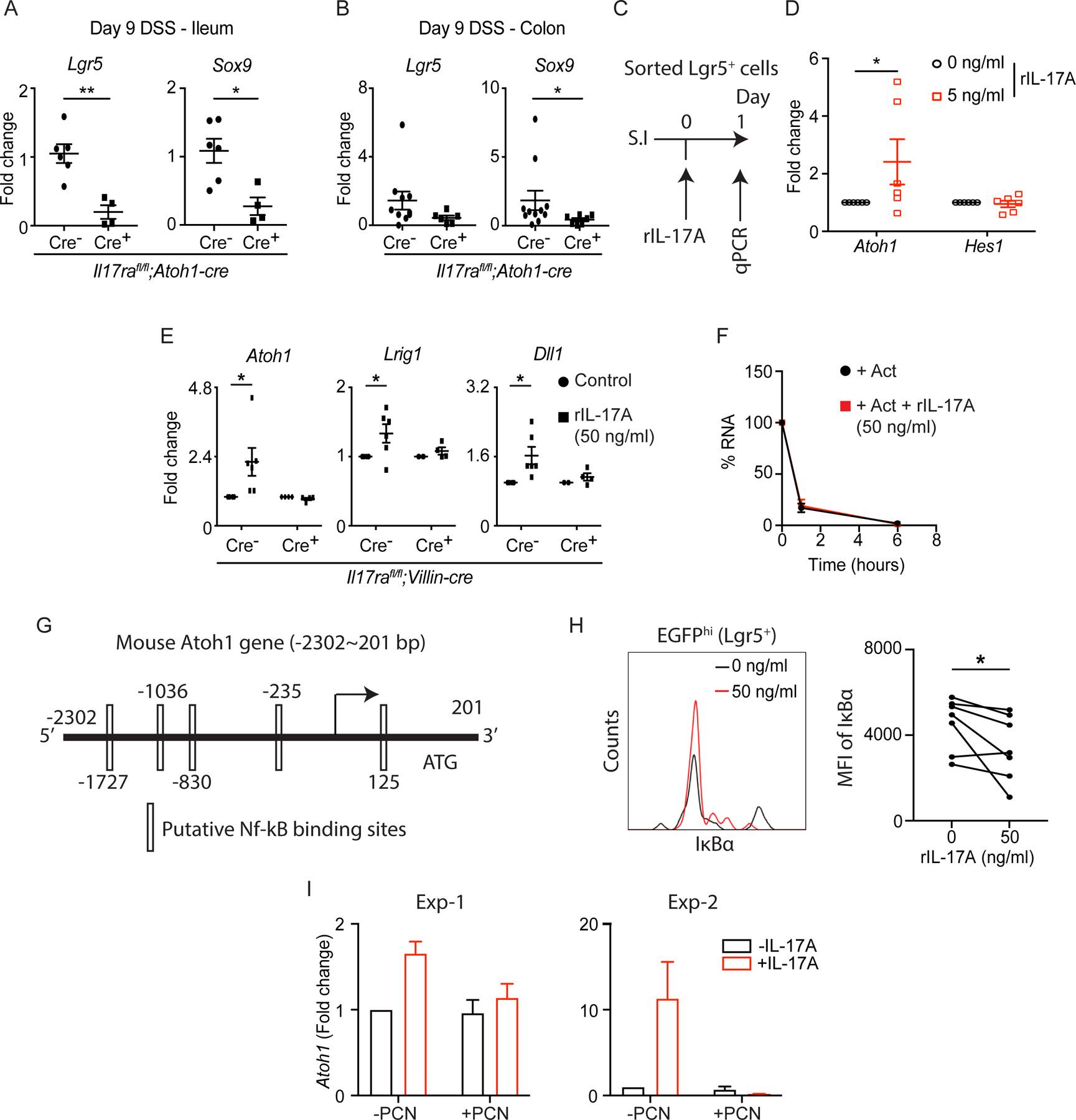
Figure 5. IL-17A signaling in Lgr5+ ISCs regulates ATOH1 activity in an NF-κB-dependent manner.

. A-B) Il17rafl/fl;Atoh1-cre mice were treated with 2.5% DSS for 8 continuous days followed by 1 day of water. RT-PCR data depict the expression of Lgr5 and Sox9 in the terminal ileum (A) and distal colon (B) harvested on day 9. C-D) Crypts were isolated from the ileum of Lgr5-EGFP-creERT2+ mice. EGFPhi cells (Lgr5+ ISCs) were sorted by fluorescence-activated cell sorting (FACS). The schematic diagram shows the strategy of IL-17A treatment on sorted Lgr5+ ISCs (C). The expression of Atoh1 and Hes1 was analyzed by RT-PCR (D). E) Crypts isolated from the ileum of Il17rafl/fl;Villin-cre mice were used for organoid culture under recombinant IL-17A (50 ng/ml) treatment. The expression of Atoh1, Lrig1 and Dll1 was analyzed by RT-PCR. F) Primary C57BL/6J organoids were treated with actinomycin D (5 μg/mL) and recombinant IL-17A (50 ng/ml). Atoh1 transcripts were quantified at the indicated time points by RT-PCR. G) Putative NF-κB binding motifs are predicted in the mouse Atoh1 promoter. H-I) Crypts were isolated from the ileum of Il17rafl/fl;Lgr5-EGFP-creERT2+ mice (without tamoxifen administration) and used for primary organoid culture under the treatment of recombinant IL-17A (50 ng/ml) or piceatannol (10 μM). After 5 days of recombinant IL-17A treatment, organoids were processed for flow cytometry. Live Lgr5hi cells were gated and the MFI of IκBα was plotted (H). The expression of Atoh1 in organoids harvested after 5-day treatment of piceatannol was analyzed by RT-PCR (I).
Figures 5A, 5B, 5D, 5E, 5H and 5I were generated from 2–3 independent experiments. Figure 5F was generated from 3 mice in each group. Data are presented as mean ± SEM in all graphs. *P ≤ 0.05; **P ≤ 0.01 (Mann-Whitney test, two-tailed in A and B, Two-Way ANOVA in D-F, Wilcoxon matched-pairs test in H). See also Figures S4 and S5.
