
Figure 2.
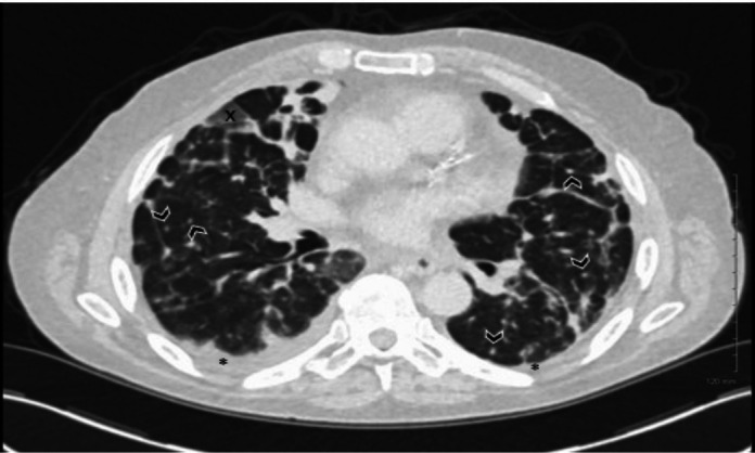
Selected axial image demonstrating widespread bilateral pulmonary nodules (black chevrons), another finding typical of LC. bilateral pleural effusions again demonstrate (black asterisks). Ground glass opacification which was visualised in a scattered distribution throughout the lungs (black figure X) was thought to most likely represent superimposed infection and is unrelated to the LC.