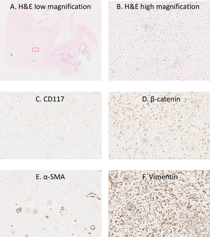
Figure 6.
Histopathological images of desmoid tumour. (A) H&E low-magnfiication (1x) of whole slide with bowel on bottom right hand side and highlighted red box for higher power magnification of subsequent slides. (B) H&E high-magnification (40x) of highlighted area in (A) showing pale eosinophilic cytoplasm with no cytological atypia. (C) Immunostaining demonstrating no staining for CD117. (D) Immunostaining showing focal beta-catenin staining. (E) Immunostaining showing focal alpha smooth muscle actin staining. (F) Immunostaining showing intense vimentin staining.

