Figure 1.
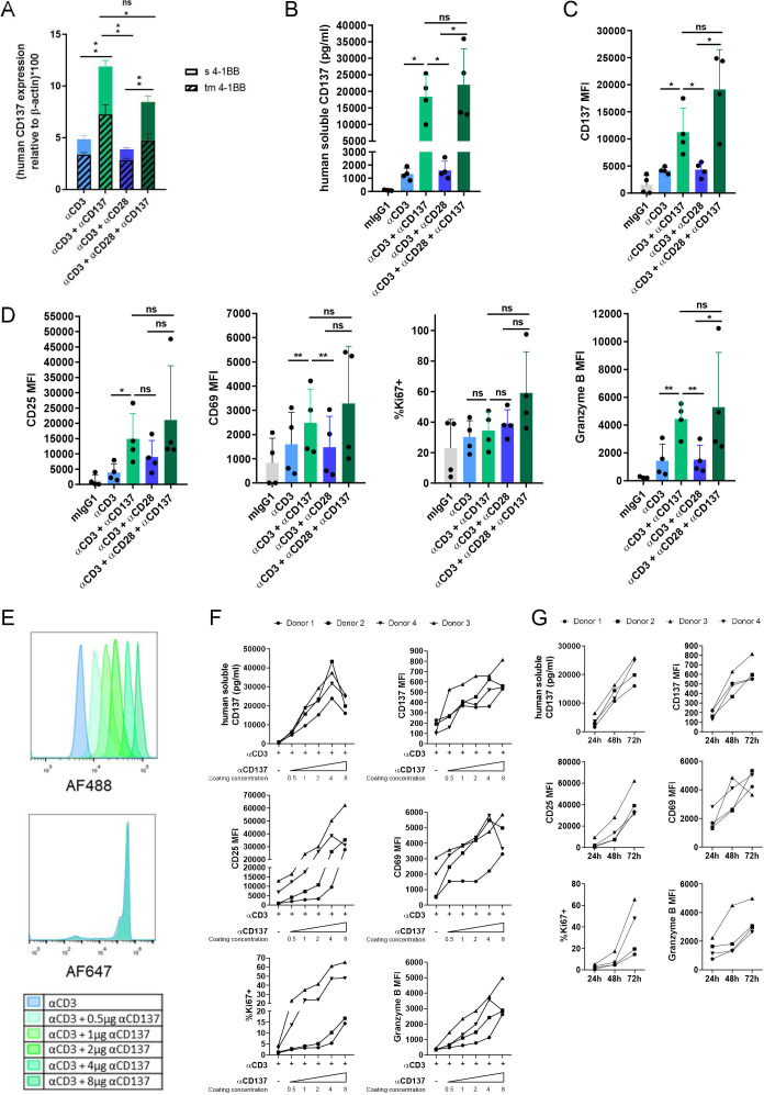
Soluble and transmembrane CD137 are upregulated by CD137 costimulation. (A) Quantitative RT-PCR measurements of mRNAs encoding transmembrane and sCD137 in isolated human CD8 T cells from three different donors stimulated by anti-CD3ε mAb bound to the culture plate in conjunction or not with anti-CD137 or anti-CD28 as indicated. (B) Measurement of the sCD137 concentration in the 48 hours tissue culture supernatants. (C) Surface expression levels of CD137 on CD8 T lymphocytes stimulated as in part A. (D) Surface expression levels of CD25 and CD69 as well as intracellular staining levels of Ki67 and Granzyme B in experiments as in part B. (E) Microbeads were covalently cocoated with fixed concentrations of anti-CD3ε tagged with AlexaFluor647 and serially increasing concentrations of anti-CD137 mAb tagged with AlexaFluor488 (0.5–8 µg of mAb per 50 µL of beads). Flow cytometry was used to show levels of microbead coating by relative fluorescence intensity units. F shows stimulation of CD8 T cells isolated from four healthy donors cocultured with the microbeads as indicated measuring sCD137 in culture supernatants or the intensity of surface expression of CD137, as well as other markers of T cell activation as indicated. Results were assessed at 72 hours of culture. G shows a time-course of these parameters in the cultures using the beads coated with the highest content of anti-CD137. Bars indicate mean±SEM. Experiments were performed with four independent donors. Statistical significance was assessed by Student’s paired t-test in parts A, B, C and D, and Wilcoxon test in part D (CD25 MFI). *p<0.05, **p<0.01. MFI, mean fluorescence intensity; ns, not significant.

