Figure 2.

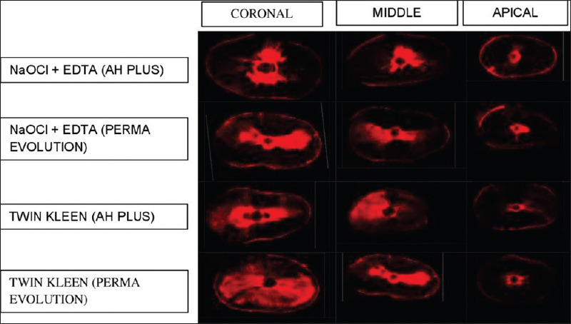
A representative confocal laser scanning microscopy image of a sample from each group at 8 mm, 5 mm, and 2 mm levels as coronal, middle, and apical third regions of root canal, respectively

A representative confocal laser scanning microscopy image of a sample from each group at 8 mm, 5 mm, and 2 mm levels as coronal, middle, and apical third regions of root canal, respectively