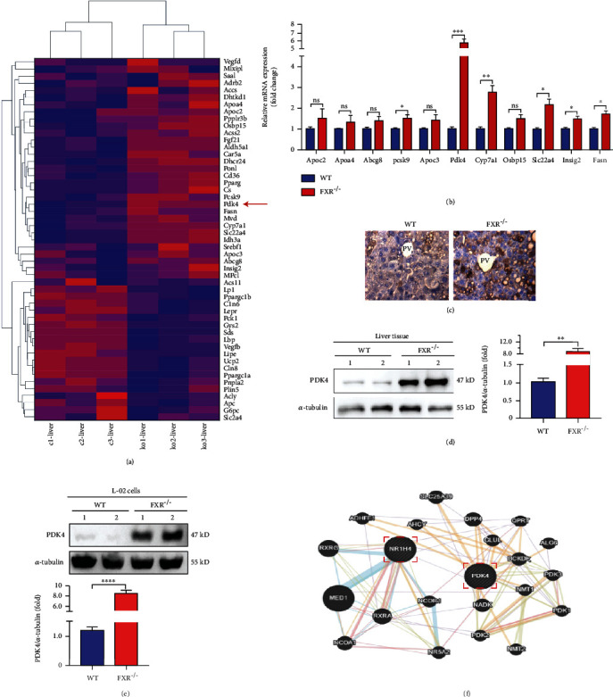
Figure 3.

FXR deficiency increased hepatocyte's expression of PDK4. (a) An unbiased RNA-seq expression profiling heat map between liver tissues of WT littermates (c1, c2, and c3) and FXR-null mice (ko1, ko2, and ko3). (b) mRNA levels of PDK4 and genes related to lipid metabolism were detected by qRT-PCR in the liver of FXR-null and WT mice, n = 3. (c) Immunohistochemical staining has detected the expression of PDK4 in liver tissue, bar = 50 μm. Representative images of liver tissues of control (WT) and FXR null mice with immunohistochemical staining are shown, n = 3. (d) Liver tissue lysates were immunodetected for PDK4 and α-tubulin, relative levels of PDK4/α-tub are shown, and two independent FXR-null mice (KO1 and 2) were analyzed, n = 3. (e) FXR-null L-02 (KO) and control cells (WT) lysates were immunodetected for PDK4 and α-tubulin, relative levels of PDK4/α-tub are shown, two independent FXR-null L-02 cell lines (KO1 and KO2) were analyzed. (f) Through the predictive website of genetic interactions-Gene MANIA (http://genemania.org), searched for links between FXR and PDK4. ∗P < 0.05, ∗∗P < 0.01, ∗∗∗P < 0.001, ∗∗∗∗P < 0.0001, n = 3.
