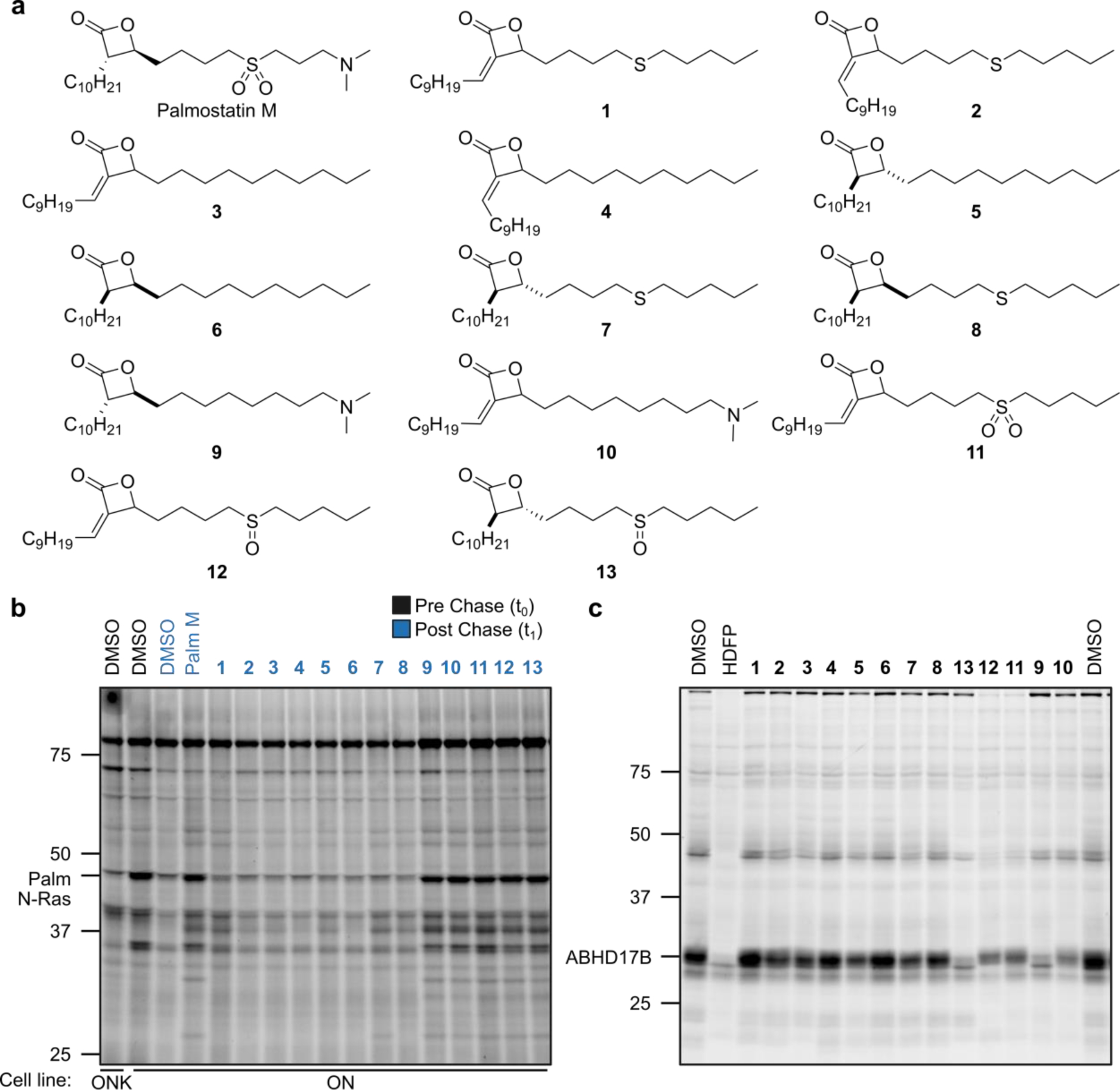
Figure 1. Structures and initial screening of palmostatin analogs.

a, Structures of Palmostatin M (Palm M) and analogs. b, Effect of palmostatin analogs on dynamic protein palmitoylation as detected by pulse-chase metabolic labeling of ON and ONK cells with the 17-ODYA probe followed by CuAAC conjugation to a Rh-N3 reporter tag, SDS-PAGE, and in-gel fluorescence scanning. Compounds were screened at 20 μM (Palm M control screened at 10 μM). c, ABHD17 inhibitory activity of palmostatin analogs measured by gel ABPP of HEK293T cells stably expressing ABHD17B. Compounds, including HDFP, were screened at 20 μM and incubated with cells in situ for 4 h prior to lysis, treatment with FP-Rh (1 μM, 1 h), SDS-PAGE, and in-gel fluorescence scanning. The screens in b and c were performed once. Note that the order of compounds 9–13 is differently arranged in b and c.
