Fig. 1.
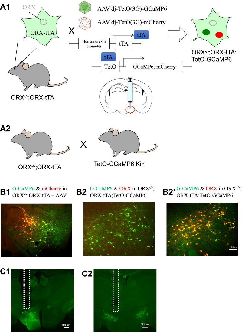
Animal models for measuring putative orexin neuronal activity. A1 Schematic drawing showing specific expression of GCaMP6 and mCherry in putative orexin neurons by injecting adeno-associated virus (AAV) vectors into the hypothalamus of preproorexin knockout (ORX−/−) and orexin-tTA transgenic (ORX-tTA) double-mutant mice. We refer to this animal as “model 1” in this study. A2 Schematic drawing showing specific expression of GCaMP6 in putative orexin neurons by crossing double mutants of ORX−/− and ORX-tTA with TetO-GCaMP knock-in mice. We call this animal “model 2” in this study. B1 Histological confirmation showing overlapping distribution of GCaMP6 (green) and mCherry (red) in the hypothalamus of a model 1 mouse. B2 Histological confirmation showing GCaMP6 (green) in the hypothalamus of a model 2 mouse. Note that no orexin-like immunoreactivity was found, as expected. B2′ Histological confirmation showing overlapping distribution of GCaMP6 (green) and orexin-like immunoreactivity (red) in the hypothalamus of a mouse carrying the orexin-knockout heterozygous allele in model 2. C1 & C2 Confirmation of fiber tracts after fluorescent recordings. The dashed line indicates the location of the inserted optic fibers. The left panel C1 shows a brain from a model 1 mouse and the right panel C2 shows a brain from a model 2 mouse

