Figure 4.
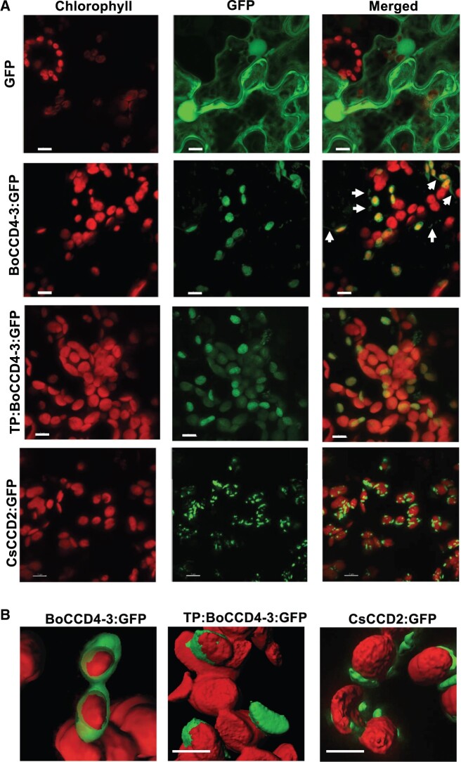
Subcellular localization of BoCCD4-3 in N. benthamiana leaves. A, Confocal images of N. benthamiana leaves expressing (top to bottom): GFP, BoCCD4c:GFP, TP:BoCCD4c:GFP (BoCCD4c fused to the CsCCD2 transit peptide), and CsCCD2:GFP fusion proteins. Red (chlorophyll fluorescence), green (GFP fluorescence), and merged (overlap of chlorophyll and GFP fluorescence) are shown. The unfused GFP protein shows the typical cytoplasmic and nuclear localization. Both BoCCD4-3:GFP and CsCCD2:GFP localize to plastids. Scale bars: 7 μm. Arrows in the BoCCD4c:GFP composite image point at green fluorescent protrusions of the plastid stroma (stromules). B, 3D reconstruction of red and green fluorescence in plastids expressing BoCCD4-3:GFP, TP:BoCCD4-3:GFP, and CsCCD2:GFP. The latter localizes to plastid-associated speckles (Demurtas et al., 2018). Scale bars: 7 μm.

