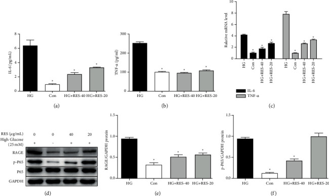
Figure 4.

RES's effects of on HG-stimulated activation of the RAGE/NF-κB and inflammatory cytokines production. H9c2 cells were subjected to treatment with control medium or suggested concentrations of RES (20 and 40 μg/mL) for 24 h. ELISA, RT-PCR, and WB analysis of IL-6, TNF-α, RAGE, p-NF-κB P65, P65, and GAPDH levels were performed. (a) IL-6's secretion level of IL-6 in supernatant; (b) TNF-α's secretion level in supernatant; (c) IL-6 and TNF-α's mRNA levels in H9c2 cells; (d) representative WB images of RAGE, p-P65, P65, and GAPDH; (e) RAGE's protein expression; (f) p-P65's protein expression. The levels of protein were determined by densitometry and normalised to the level of GAPDH. We normalised the relative mRNA levels to that of GAPDH. The data are presented as the mean ± SD of the three repeated trials; ∗p < 0.05vs. HG group.
