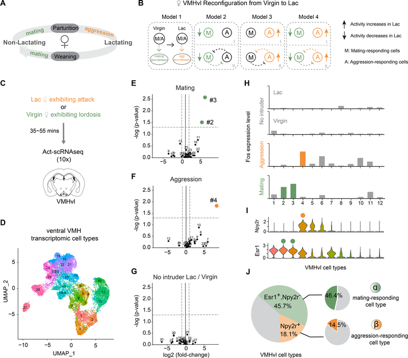
Figure 1.
Distinct VMH transcriptomic cell types are activated during female mating and aggression.
(A) Female mice adjust social behaviors according to their lactation cycle. The start of the lactating phase is always accompanied by a decrease in sexual behaviors and an onset of aggressive behaviors; at the end of the lactating phase, sexual behaviors resume, and aggression disappears.
(B) Potential cellular mechanisms underlying changes in female VMHvlEsr1 activity from the virgin to the lactating state. Model 1: The VMHvlEsr1 neurons controlling socio-sexual behavior are the same in virgin and lactating females, and critical changes occur at their downstream targets to promote mating (in virgins) or aggression (in lactating dams). (In a variant of Model 1 (not shown) different cells control mating vs. aggression, but their state-dependent behavioral output is determined by changes in their downstream targets.) Models 2–4, distinct subsets VMHvlEsr1 cells control mating in virgins and aggression in lactating dams. Their relative activity differs depending on the animal’s state and determines their behavioral output. The changes in lactating females could involve a decrease in the activity of mating neurons (Model 2); an increase in the activity of aggression neurons (Model 3); or both (Model 4). Models 2–4 can be further subdivided according to the presence (II) or absence (I) of reciprocal inhibitory interactions (likely indirect) between mating and aggression cells. Colored symbols indicate where the lactation-dependent changes occur. The change in the activity of these different subpopulations could reflect cell-intrinsic changes, or changes in input strength.
(C) Schematic of Act-seq protocol.
(D) UMAP plot color-coded by clusters identified in tissue dissected from female ventral VMH (N=19,103; 27 clusters).
(E-G) “Volcano plots” showing Fos expression levels in 27 VMH cell types from the following animals: (E) lactating females exhibiting attacks vs. control, (F) virgin females exhibiting lordosis vs. control, (G) control lactating females vs. control virgin females. Colored dots indicate cell types with Fos expression fold change >2 (x axis cut-off) and P <0.05 (y axis cut-off; gray dashed lines).
(H) Fos expression levels in 12 VMHvl cell types from control lactating females, control virgin females (No intruder), lactating females exhibiting attacks (Aggression) and virgin females exhibiting lordosis (Mating). Colored bars show T-types with statistically significant increases (fold change >2 & P <0.05) in Fos expression relative to no-intruder controls (see also Figure S2C).
(I) Violin plot illustrating expression levels of Npy2r and Esr1 in 12 VMHvl cell types.
(J) Summary of composition of VMHvl cell types. Left, shaded green: VMHvlEsr1+,Npy2r− cells; shaded yellow: VMHvlNpy2r+ cells; gray: VMHvlEsr1−,Npy2r− cells. Right, green: Percentage of mating-activated cell types (#2, #3), as detected by a significant and >2-fold increase in Fos expression, among VMHvlEsr1+,Npy2r− cells (α); orange: Percentage of aggression-activated cell type (#4) as detected by Fos expression among VMHvlNpy2r+ cells (β). Fos expression in VMHvl substantially underestimates the fraction of neurons active during a given behavior, as measured by electrophysiological recordings (Lin et al., 2011).

