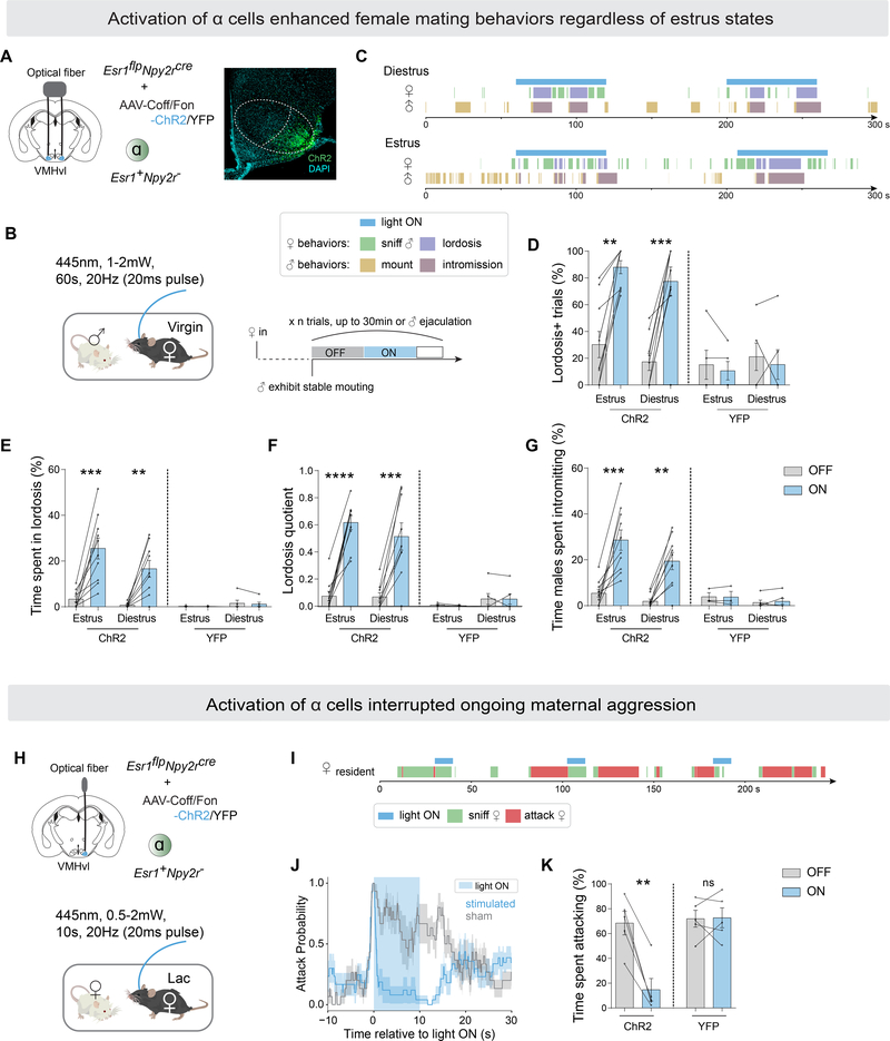
Figure 2.
VMHvl α cells control female receptivity and inhibit maternal aggression.
(A) Left, strategy to activate VMHvlEsr1+,Npy2r− cells (α) in virgin females by optogenetics. Right, representative ChR2 expression in VMHvl α cells. Scalebar, 200um.
(B) Behavioral paradigm and illustration of stimulation scheme for one session (also see Methods).
(C) Representative raster plots illustrating light-induced behaviors in tested female and paired male.
(D) Fraction of trials where female exhibited lordosis,
(E) fraction of time female spent in lordosis,
(F) lordosis quotient (lordosis time/male mounting or intromission time),
(G) fraction of time males spent intromitting during light ON or light OFF periods, in estrus or diestrus intact (non-ovariectomized) ChR2/YFP-expressing females.
(H) Strategy to activate VMHvlEsr1+,Npy2r− cells (α) in lactating females by optogenetics.
(I) Representative raster plots illustrating light-induced behaviors in aggressive lactating female.
(J) Average attack probability. In blue, stimulated trials with light ON; in grey, sham trials with light OFF.
(K) Fraction of time lactating female spent attacking during 10-second stimulated or sham periods.
*p<0.05; **p<0.01; ***p<0.001; ****p<0.0001. Mean ± SEM.

