Figure 5.
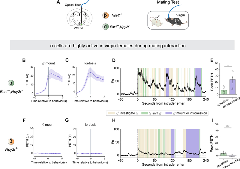
VMHvl α cells are highly active in virgin females during mating behaviors.
(A) Schematic illustrating fiber photometry recording in VMHvl and behavioral test in virgin females.
(B-C) PETH of GCaMP6m fluorescence in α cells aligned to onset of (B) male mounting and (C) lordosis.
(F-G) PETH in β aligned to onset of (F) male mounting and (G) lordosis.
(D, H) Representative normalized calcium traces from (D) α cells and (H) β cells during mating interaction. Colored shading marks behavioral episodes.
(E, I) Peak of PETH from (E) α cells and (I) β cells aligned to onset of appetitive phase (sniff) or consummatory phase (male mounting or intromission) during mating interaction.
*p <0.05; ns, not significant. Mean ± SEM.

