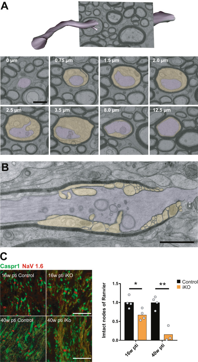
Fig. 6. Juxtaparanodal myelin tubulation and loss of nodal organization.
A Segmentation of axon and myelin tubules in an image stack acquired by FIB-SEM in the optic nerve of iKO 26 weeks pti (shown in Supplementary Movie 2). The distance along the internode is indicated in the images. Membrane tubules emerge at the juxtaparanode (0.75–3.5 µm) while most of the internode is unaffected. B Longitudinal TEM section reveals the juxtaparanode localization of the tubules and the detachment of the paranodal loops. This phenotype was observed independently in two groups of female mice (n = 7 controls; n = 10 iKOs and n = 3 controls; n = 5 iKO) and two groups of male mice (n = 3 controls; n = 5 iKOs and n = 2 controls; n = 4 iKOs). C Confocal light microscopy of immunofluorescence staining of the nodal marker NaV1.6 and paranodal marker Caspr1 on optic nerve cryosections reveals loss of functional nodes of Ranvier (two-tailed unpaired t test, control vs iKO; 16 weeks pti: p = 0.0207; 40 weeks pti: p = 0.0051 (p < 0.05 (*), p < 0.01 (**)). Source data are provided with this paper. Scale bars: 500 nm (A, B), 10 µm (C).

