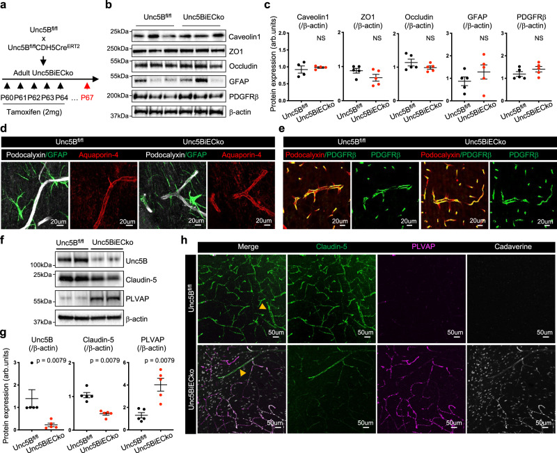
Fig. 2. Unc5B controls Claudin-5 and PLVAP expression.
a Unc5B gene deletion strategy using tamoxifen injection in adult mice. Western blot (b) and quantification (c) of brain protein extracts at P67. Quantification of Caveolin-1 expression was performed on n = 4 Unc5Bfl/fl and n = 4 Unc5BiECko brains. Quantification of ZO1, Occludin, GFAP and PDGFRβ expression was performed on n = 5 Unc5Bfl/fl and n = 5 Unc5BiECko brains. Each dot represents one mouse. One control mouse was set as 1. d, e Immunofluorescence staining with the indicated markers and confocal imaging of brain sections, reproduced on n = 4 Unc5Bfl/fl and n = 4 Unc5BiECko brains. Western blot (f) and quantification (g) of P67 brain protein extracts, n = 5 Unc5Bfl/fl and n = 5 Unc5BiECko brains. Each dot represents one mouse. One control mouse value was set as 1. h Immunofluorescence staining with the indicated antibodies and confocal imaging of P67 piriform cortex 30 min after i.v cadaverine injection, reproduced on n = 4 Unc5Bfl/fl and n = 4 Unc5BiECko brains. Arrowheads: larger vessels. All data are shown as mean ± SEM. NS non-significant. Two-sided Mann–Whitney U test was performed for statistical analysis. Source data are provided as a Source Data file.

