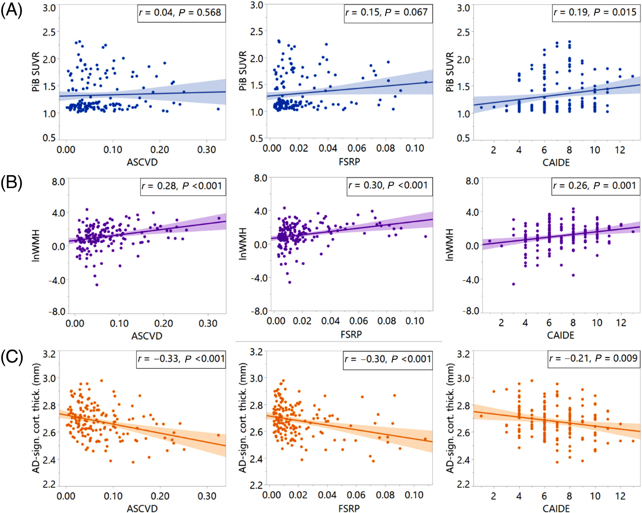
FIGURE 1.

Vascular risk factor (VRF)-imaging scatterplots. VRFs (x-axes) versus (A) Global PiB SUVR, (B) log-norm. WMH volume, and (C) temporal lobe cortical thickness (y-axes). ASCVD, atherosclerotic cardiovascular disease risk estimate from the pooled cohort equation; FSRP, Framingham stroke risk profile; CAIDE, cardiovascular risk factors, aging and incidence of dementia risk score; PiB SUVR, Pittsburgh compound B Standardized Uptake Value Ratio; lnWMH, log-normalized white matter hyperintensity volume; AD-sign. cort. thick., temporal lobe cortical thickness
