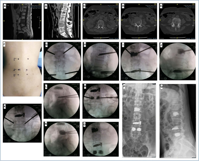
Fig. 5.
Personalized percutaneous vertebroplasty (PVP) procedure. A and B Preoperative sagittal MRI and CT scans show L4 and L5 involvement (low signal on T1WI and slightly high signal on T2WI); C–E preoperative transverse CT scans show L2, L4, and L5 involvements; F preoperative body surface location of L2, L4, and L5; G and H C-arm fluoroscopy shows that puncture needles are located in L2 and L4 vertebrae; I and J bone cement is injected into the vertebral bodies of L2 and L4; K and L C-arm fluoroscopy shows that puncture needles are located in L5 vertebra; M and N bone cement is injected into L5; O C-arm fluoroscopy shows good dispersion of bone cement in L2, L4, and L5; P and Q postoperative X-ray shows a good dispersion of bone cement in L2, L4, and L5

