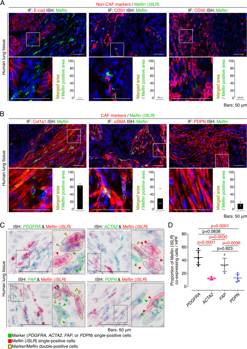
Figure S2. Meflin is expressed in stromal fibroblasts but not in other types of cell in human non-small cell lung cancer (NSCLC).
(A) Tissue sections prepared from a human NSCLC sample were stained for the epithelial cell marker E-cadherin, the endothelial cell marker CD31, and the leukocyte marker leukocyte common antigen by immunofluorescence (IF) (red) and Meflin mRNA by ISH using RNAscope technology (green). Note that no Meflin+ cells were positive for E-cadherin, CD31, and leukocyte common antigen. The graphs in the insets show the ratios of the merged areas to those of the Meflin+ areas. (B) Tissue sections prepared from a human NSCLC sample were stained for the cancer-associated fibroblast (CAF) markers Col1a1, α-smooth muscle actin, and podoplanin by IF (red) with Meflin mRNA detection by ISH using RNAscope technology (green). The graphs in the insets indicate the ratios of the merged areas to those of the Meflin+ areas. (C) Higher magnified images of duplex ISH for Meflin (red) and other CAF markers (green: PDGFRA, ACTA2, FAP, or PDPN) in CAFs of human NSCLC (related to Fig 1F). Green, yellow, and red arrowheads denote cells that are single-positive for the indicated CAF markers, double-positive for the indicated CAF markers and Meflin, and single-positive for Meflin, respectively. Boxed regions were further magnified in adjacent panels. (D) The quantification and comparison of Meflin+ cells across CAFs positive for the indicated CAF markers. All CAFs that were positive for respective ISH signals in five HPFs were evaluated and quantified.

