
FIGURE 2.
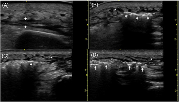
Point‐of‐care ultrasound of the affected right tibia in a transverse (A) plane demonstrating perifascial fluid accumulation (between plus symbols). Point‐of‐care ultrasound of the affected right foot in transverse (B,C) and longitudinal (D) planes demonstrating hyperechoic air foci (arrows) with dirty posterior shadowing representative of necrotizing fasciitis. Subcutaneous edema (asterisks) clinically consistent with overlying cellulitis