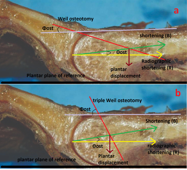
Figure 6. Weil osteotomy (A) and triple Weil osteotomy (B).
The first panel shows a Weil osteotomy, where the proximal displacement of the head fragment is calculated using the most distal edge of the proximal fragment as a reference. The second panel shows triple Weil osteotomy, where the proximal displacement of the metatarsal head is measured by the thickness of the bone fragment removed by the second cut. In both cases, the shortening of the metatarsal (B) is along its longitudinal axis. The radiographic shortening (R) is along the plane of reference. Θost: osteotomy angle.

