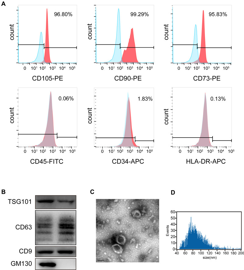
Figure 1.
Characterization of iMSC and iMSC-sEVs. (A) Surface antigen profile of iMSC evaluated by flow cytometry. (B) Western blotting showing the expression of exosomal markers including CD9, TSG101, and CD63 in iMSC-sEVs, but not the negative marker GM130. (C) Representative image of iMSC-sEVs observed by TEM. Scale bar = 100 nm. (D) Particle size distribution of iMSC-sEVs measured by nano-flow cytometer.

