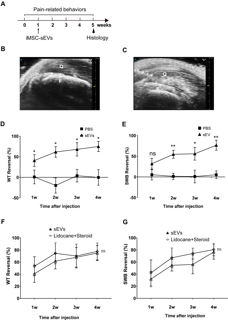
Figure 2.
Analgesic Effect of iMSC-sEVs on Rat Tendinopathy Model. (A) Diagram showing timing for the model establishment and iMSC-sEVs treatment. Pain-related behaviors were assessed once per week for 5 weeks and histopathological changes were evaluated at 5 weeks after model establishment. (B) Ultrasonogram of rat’s right leg (anterior view) around the quadriceps tendon showing quadriceps (Q) and femur (F). (C) Ultrasonogram showing the relative position of needle (white triangles) used to inject 4% carrageenan (100 μL bolus) solution around the quadriceps tendon. Asterisks (white) in (B and C) indicate the position of the quadriceps tendon. Pain-related behaviors were performed by using PWT (D) and SWB (E) reversal (%) up to 4 weeks after tendinopathy model establishment. PWT (F) and SWB (G) reversal (%) up to 4 weeks after tendinopathy model establishment. N = 5 rats for each group. SWB = static weight bearing; PWT = hind-paw withdrawal threshold; Data are presented as mean ± SD. *P < 0.05. **P < 0.01. ns indicates no significant difference.

