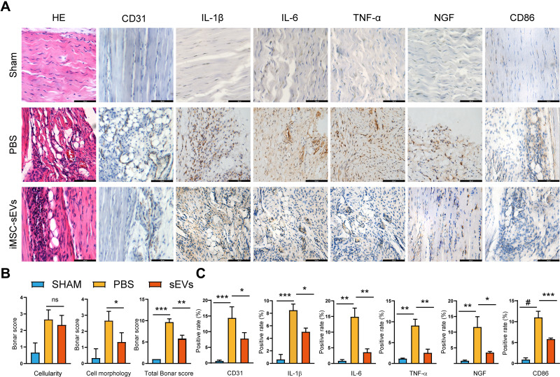
Figure 3.
iMSC-sEVs alleviate inflammatory cytokine infiltration in rat tendinopathy model. (A) Representative photomicrographs of H&E and immunohistochemically stained tissue sections of different groups at week 5 after model establishment. Positive immunostaining of IL-1β, IL-6, TNF-α, and NGF was visualized with DAB (brown), and nuclei were counterstained with hematoxylin (blue). Scale bar = 100μm. (B) Modified Bonar score, including cell morphology and cellularity, is used for semiquantitative histology analysis. C Quantitative analysis of immunohistochemical staining. Data are expressed as mean ± SD. *P < 0.05. **P < 0.01. ***P < 0.001. #P < 0.0001.

