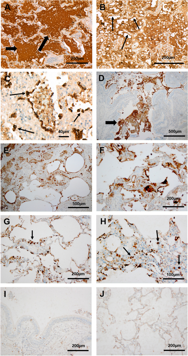
Fig. 1.

Pulmonary DMBT1 expression in patients with CF and corresponding controls. Immunohistochemical analysis of DMBT1 expression in post-mortem lung tissue of a patient with cystic fibrosis (A–C), in explanted lungs of CF patients due to lung transplantation (D–H) and in corresponding controls without lung disease (I, J). DMBT1 localization is displayed as brown staining. A The alveoli and small airways contained luminal mucus with intense DMBT1 staining (arrows). Magnification: × 10. B The respiratory epithelial cells (small arrow) showed high expression of DMBT1. Magnification: × 15. C Multiple DMBT1-positive macrophages (arrows) were observed in the aveoli and small airways. Magnification: × 40. D–F The small airways (D, arrow) and alveoli (E, F) showed mucus stained with immunohistochemistry using an antibody against DMBT1. Magnification: × 4. G, H DMBT1-positive macrophages (arrows) were visible in the aveoli (small arrows). Magnification: × 10 (G) and × 20 (H). I, J Control tissue without lung disease showed only distinct DMBT1 expression compared to pulmonary DMBT1-expression in CF patients. Magnification: × 10
