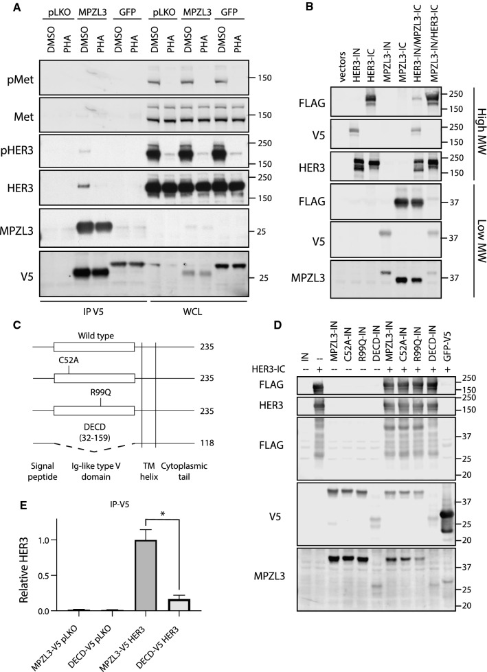
Fig. 5.
HER3 interacts with MPZL3. a Co-immuno-precipitation analysis of KatoII cells stably transduced with MPZL3- or GFP-V5 expression vectors, or control (pLKO). Cells were treated with PHA-665752 (0.5 mM, 1 h) or with vehicle control (DMSO). Immunoprecipitation (IP), Whole cell lysate (WCL). b HER3 and MPZL3 were analyzed by split intein-mediated protein ligation (SIMPL) assay to measure direct protein–protein interaction. c Schematic of MPZL3 mutant constructs. d FLAG transfer by point mutants and the ΔECD mutant were compared to wild-type MPZL3 using the SIMPL assay. e Co-immunoprecipitation of MPZL3 and HER3 was quantified, comparing HER3 recovered by the full-length or ΔECD MPZL3 mutants. Data are means (bars) of three individual replicates. Student’s t test; *p ≤ 0.05; **p ≤ 0.01; ***p ≤ 0.001; ****p ≤ 0.0001. Error bars indicate SEM

