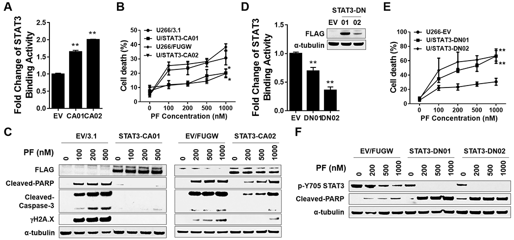
Figure 6. STAT3 inhibition by Chk1 inhibitor through a STAT3-dependent manner.

A-C, U266 were transfected with pcDNA3.1 (empty vector) or pcDNA-STAT3 CA (FLAG fusion), and STAT3-CA01 was selected. STAT3-CA02 was selected in U266 cells infected with a lentivirus harboring STAT3 CA (FLAG fusion). A, The STAT3 DNA-binding ELISA was used to evaluate STAT3 activity in STAT3-CA cell. B, cells were exposed (24 hr) to indicated concentrations of PF, followed by flow cytometry to monitor the percentage of apoptotic (7-AAD+) cells. Values represent the means ± S.D. for three experiments performed in triplicate. * = P < 0.05; ** = P < 0.01. C, Western blot analysis of FLAG, cleaved-PARP, cleaved-Caspase-3, and γH2A.X was performed. α-tubulin controls were assayed to ensure equivalent loading and transfer. D-F, U266 cells infected with a lentivirus harboring STAT3 DN (FLAG fusion). STAT3-CA01/02 were selected. Assays were performed as in A-C, ** = P < 0.01.
