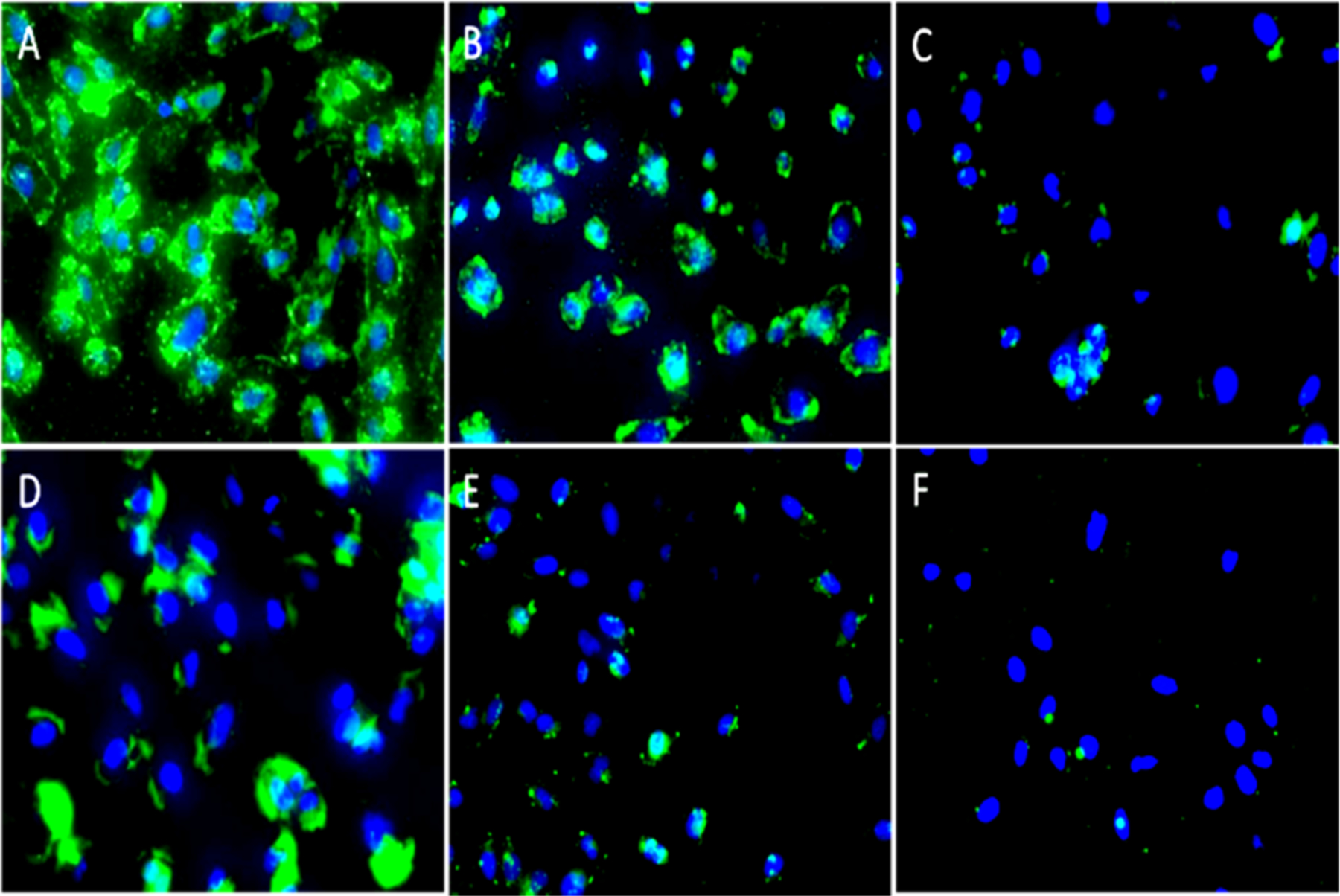
Figure 5.

Fluorescence images of the D556 medulloblastoma cells treated with different nanoparticle conjugates. Upper panels: Cells treated with nanorod conjugates Tf-IONRs (A) and BSA-IONRs (B) and cells pretreated with a fixed amount of Tf, followed by treatment with Tf-IONRs (C, blocking control experiment). Lower panel: Cells treated with spherical Tf-IONPs (D); cells treated with spherical BSA-IONPs (E); and cells pretreated with a fixed amount of Tf, followed by treatment with Tf-IONPs (F, blocking control experiment). Magnification: 20×. FITC was used to label Tf or BSA. DAPI (blue) was used to stain cell nuclei.
